Basal cell carcinoma treatment: options and what to expect
Basal cell carcinoma (BCC) is the most common form of skin cancer and typically grows slowly in areas exposed to the sun. Treatment choices depend on the size, location, subtype, and patient factors. This article outlines common approaches used by dermatology and medical teams so you can understand typical pathways and questions to discuss with your clinician.

This article is for informational purposes only and should not be considered medical advice. Please consult a qualified healthcare professional for personalized guidance and treatment.
What is basal cell skin cancer?
Basal cell carcinoma arises from basal cells in the outer layer of the skin and most often occurs on sun-exposed sites such as the face, ears, neck, and hands. Unlike melanoma, BCC rarely spreads to distant organs, but it can invade locally and cause damage to underlying tissues if left untreated. Dermatology evaluation typically includes a clinical exam and may include dermoscopy or biopsy to confirm the diagnosis and to determine subtype, such as nodular, superficial, or morpheaform (infiltrative).
Early detection is important: smaller, well-defined tumors generally have more treatment options and lower risk of cosmetic or functional impairment following treatment. Patients with a history of significant sun exposure, tanning bed use, or weakened immune systems are at higher risk and may be offered more frequent skin checks.
How is BCC diagnosed in dermatology?
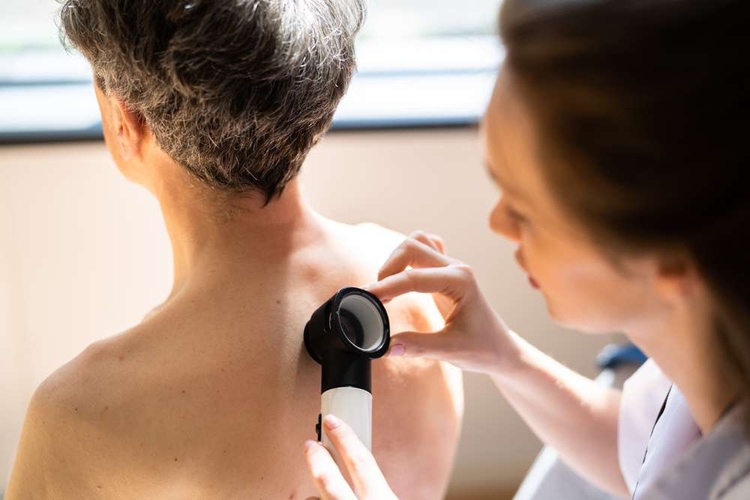
The diagnostic process usually begins with a thorough skin examination by a dermatologist or trained clinician. Suspicious lesions are assessed visually and with tools like a dermatoscope to look for characteristic features. If there is uncertainty, a small sample of the lesion (skin biopsy) is taken and examined histologically by a pathologist to confirm basal cell carcinoma and to classify its subtype.
Biopsy results guide medical decision-making: for example, superficial BCCs may respond well to topical or superficial therapies, whereas infiltrative or high-risk subtypes often require surgical removal with clear histologic margins. The pathology report will note margin status, depth, and subtype — details that are important when planning subsequent treatment.
Common medical treatment options
Non-surgical medical treatments are appropriate for select BCCs, especially small, superficial lesions or for patients who cannot have surgery. Options include topical agents such as imiquimod or 5-fluorouracil, which stimulate local immune response or interfere with cancerous cell growth. Photodynamic therapy combines a photosensitizing agent with light exposure to selectively destroy abnormal cells and may be used for superficial lesions.
Systemic medical therapies are reserved for advanced, recurrent, or metastatic disease and include targeted agents (for example, hedgehog pathway inhibitors) under specialist supervision. These systemic medications have specific side effect profiles and require monitoring by oncology or dermatology teams. The choice of medical treatment is individualized and balances efficacy, side effects, cosmetic outcomes, and patient preferences.
Surgical and procedural treatments
Surgical removal is the most common and often curative approach for many BCCs. Standard excision involves removing the tumor with a rim of normal-appearing skin; the specimen is then examined to ensure clear margins. Mohs micrographic surgery is a tissue-sparing technique performed by specially trained surgeons where margins are examined layer by layer during the procedure, offering low recurrence rates and preservation of healthy tissue—particularly useful for lesions on the face or in functionally sensitive areas.
Other procedural options include curettage and electrodessication for small, low-risk lesions, or cryotherapy in select superficial cases. For larger defects after surgery, reconstructive techniques ranging from primary closure to skin grafts and local flaps may be necessary. The chosen procedure depends on tumor characteristics, cosmetic considerations, and local services available in your area.
Follow-up care and prevention
After treatment, regular follow-up is recommended to monitor for recurrence and to screen for new skin cancers, since a history of BCC increases the risk of subsequent lesions. Follow-up frequency varies with risk but often involves skin checks every 3–12 months initially, then annually. Patients should be educated on self-examination and sun-protective behaviors, including broad-spectrum sunscreen, protective clothing, and avoidance of peak sun hours.
Prevention strategies from dermatology and public health perspectives emphasize minimizing ultraviolet exposure, treating precancerous lesions promptly, and addressing modifiable risks. For patients with multiple or recurrent BCCs, clinicians may discuss more intensive surveillance or preventive options tailored to the individual.
Conclusion
Basal cell carcinoma treatment spans dermatology assessments, biopsy-confirmed diagnosis, a range of medical and surgical therapies, and structured follow-up to reduce recurrence and preserve function and appearance. Treatment decisions are individualized based on tumor subtype, location, patient health, and preferences. Discussing risks, expected outcomes, and cosmetic implications with your medical team will help clarify the most appropriate approach for your situation.


