Clinical pathway for evaluating new-onset blurred vision in adults
A practical clinical pathway clarifies assessment steps for adults presenting with new-onset blurred vision. This summary outlines screening, focused history, targeted examinations, and escalation points to optometry or ophthalmology for further diagnostics and management.

Adults who experience new-onset blurred vision require a systematic, time-sensitive evaluation to distinguish benign refractive changes from urgent ocular or neurologic disease. Initial assessment focuses on symptom timing, monocular versus binocular presentation, associated pain, flashes, floaters, systemic symptoms, and recent medication changes. A concise, documented history guides whether immediate imaging or specialist referral is needed. Visual disturbance that progresses rapidly, is accompanied by pain, or includes neurologic signs generally warrants expedited assessment.
This article is for informational purposes only and should not be considered medical advice. Please consult a qualified healthcare professional for personalized guidance and treatment.
What are common ocular symptoms and their significance?
New blurred vision often presents alongside other ocular symptoms such as glare, halos, photophobia, floaters, or ocular discomfort. Monocular blurring typically indicates anterior segment or refractive issues involving the cornea, lens, or retina, while binocular blur often reflects refractive error or accommodative problems. Sudden onset with flashes or a shower of floaters raises concern for retinal detachment. Gradual, progressive visual decline plus glare and haloing can suggest cataract or corneal surface disease. Documenting these symptoms helps triage urgency and the likely anatomical source of vision loss.
How is refraction and acuity assessed in initial diagnostics?
Objective and subjective refraction, along with best-corrected visual acuity testing, are cornerstone diagnostics in the outpatient pathway. Distance and near acuity measurements clarify the functional impact on eyesight. Refraction can rapidly detect common, correctable causes like uncorrected refractive error, presbyopia, or changes after systemic illness. If acuity improves significantly with pinhole testing, a refractive problem is likely. When refraction and acuity do not explain the symptom severity, further ocular imaging and targeted examination become necessary.
When should ophthalmology or optometry be involved in evaluation?
Primary care clinicians and optometrists often perform initial screening and refraction. Escalation to ophthalmology is recommended for suspected retinal pathology, acute painful eye conditions, severe vision loss, or when specialized interventions (surgical, intravitreal therapy) may be required. Optometry offers comprehensive refractive and anterior segment assessment and can coordinate referrals for advanced diagnostics. Collaboration between providers ensures timely diagnostics, with ophthalmology focusing on surgical and retinal care while optometry handles many non-urgent refractive and corneal diagnoses.
How are retina and cornea causes evaluated and distinguished?
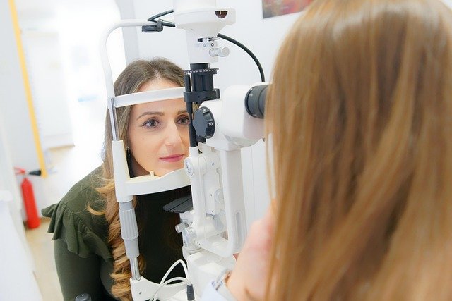
Examination of the retina and cornea uses different methods: slit-lamp biomicroscopy inspects the cornea for epithelial defects, edema, dystrophies, or infectious keratitis that cause glare and reduced acuity, while dilated fundus examination and ocular imaging assess retinal causes such as macular edema, vascular occlusion, or detachment. Optical coherence tomography (OCT) and fundus photography are common diagnostics for retina assessment; corneal topography and pachymetry help characterize corneal irregularities. The presence of pain, discharge, or surface staining points toward corneal pathology, while painless central blur or metamorphopsia often implicates the macula.
How do diagnostics address glare, imaging, and functional testing?
Diagnostics extend beyond acuity and refraction to include imaging and functional tests tailored to symptoms. OCT quantifies retinal thickness and macular pathology, visual field testing detects peripheral deficits suggestive of optic nerve or neurologic disease, and contrast sensitivity or glare testing evaluates visual function not captured by acuity alone. Corneal imaging and tear-film assessment identify surface disease causing variable vision and discomfort. Combining these tests with clinical examination improves diagnostic precision and guides whether medical therapy, optical correction, or surgical intervention is indicated.
What rehabilitation, lenses, and follow-up strategies are recommended?
Rehabilitation begins with correct prescription lenses, management of ocular surface disease, and addressing modifiable risk factors such as glycemic control in diabetes. For persistent deficits, low-vision rehabilitation and orientation resources can optimize function. Contact lenses or specialty lenses may address irregular astigmatism from corneal disease; surgical options like cataract extraction or retinal procedures are considered when structural disease explains visual loss. Follow-up intervals depend on etiology—urgent retinal conditions require immediate review, while stable refractive changes may be reassessed in weeks to months.
Clinical pathways emphasize structured history-taking, targeted physical examination, timely diagnostic testing, and clear referral thresholds to preserve eyesight and identify vision-threatening conditions promptly. A coordinated approach among primary care, optometry, and ophthalmology supports accurate diagnosis, appropriate management, and rehabilitation planning for adults with new-onset blurred vision.


