Endoscopic and Minimally Invasive Retrieval Methods Explained
Endoscopic and minimally invasive approaches are commonly used to remove urinary stones with less tissue trauma and faster recovery than traditional open surgery. This article outlines procedures such as ureteroscopy and lithotripsy and highlights how imaging and metabolic care support prevention and reduce recurrence.

Endoscopic and minimally invasive retrieval techniques aim to remove urinary stones while limiting damage to surrounding tissues and preserving renal function. These approaches span flexible and rigid ureteroscopy, percutaneous access, and various forms of lithotripsy. Patient selection, stone size and location, and imaging findings guide the choice of method. Multidisciplinary management often pairs procedural removal with metabolic and lifestyle measures to lower the risk of future stones.
This article is for informational purposes only and should not be considered medical advice. Please consult a qualified healthcare professional for personalized guidance and treatment.
How do hydration and diet affect outcomes?
Maintaining adequate hydration and attention to diet are cornerstones of both treatment planning and long-term prevention. Increased fluid intake dilutes urine and can reduce stone formation and aid passage of small fragments after procedures. Dietary adjustments—moderating sodium and balancing animal protein—interact with calcium, oxalate, and uric acid handling in the body. Clinicians often review 24-hour urine profiles to tailor recommendations that support healing after retrieval and reduce recurrence.
What role do oxalate and calcium play in recurrence?
Calcium oxalate is a common stone composition; therefore, understanding oxalate sources and calcium balance is important. Rather than restricting dietary calcium universally, many specialists advise consuming calcium with meals to bind dietary oxalate in the gut, lowering absorption. Reducing high-oxalate foods and managing sodium intake can also help. Laboratory assessment of urinary oxalate and calcium excretion guides individualized plans to lower the chance of future stones.
When is lithotripsy used and how does it work?
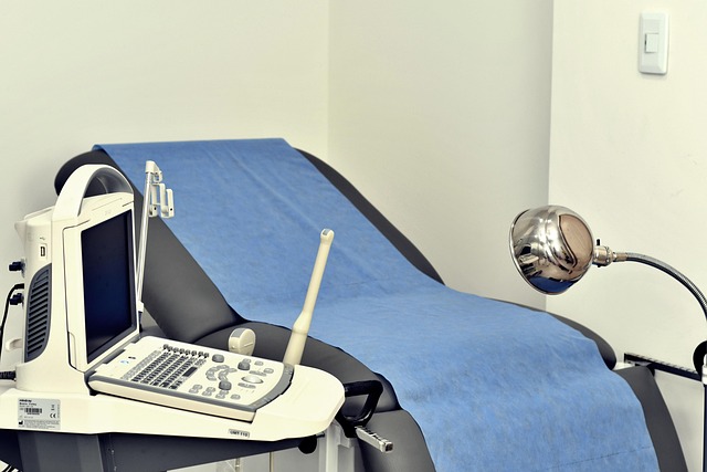
Extracorporeal shock wave lithotripsy (ESWL) and variations use focused energy waves to fragment stones so that fragments can pass naturally or be removed endoscopically. Indications depend on stone size, composition, and position in the urinary tract. Ultrasound or X-ray imaging helps localize the stone during lithotripsy sessions. While generally minimally invasive and performed on an outpatient basis, ESWL may require follow-up imaging to confirm stone clearance and to monitor for residual fragments.
What is ureteroscopy and how are stones retrieved?
Ureteroscopy involves passing a scope through the urethra and bladder into the ureter or kidney to visualize and treat stones directly. Flexible or rigid ureteroscopes allow laser lithotripsy to fragment stones, and small baskets or graspers retrieve fragments. Ureteroscopy is commonly used for stones that are not ideal for ESWL or are located in the ureter or renal collecting system. The procedure preserves anatomy and avoids an external incision, often allowing rapid recovery.
How does imaging guide procedure choice and follow-up?
Imaging—ultrasound, non-contrast CT, and fluoroscopy—determines stone size, density, and exact location, which are critical for planning a minimally invasive approach. Ultrasound is useful for screening and monitoring, while CT provides high-resolution detail about stone burden and anatomy. Post-procedure imaging confirms clearance or identifies residual fragments that might require additional intervention, and periodic imaging is part of monitoring for recurrence when clinically indicated.
How can metabolism and preventive strategies reduce recurrence?
Addressing underlying metabolic contributors—such as high urinary uric acid, low citrate, or abnormal calcium excretion—reduces recurrence risk. Metabolic evaluation may include blood tests and 24-hour urine collections to measure citrate, uric acid, calcium, and other factors tied to stone formation. Medical treatments (for example, citrate supplementation or uric acid-lowering strategies) combined with sustained hydration and dietary adjustments can decrease recurrence. Regular follow-up and tailored plans based on metabolic results support durable outcomes.
In summary, endoscopic and minimally invasive retrieval methods offer targeted options for stone removal with reduced recovery times compared with open surgery. Procedure selection relies on stone characteristics and imaging, while long-term success depends on addressing metabolic drivers and lifestyle factors such as hydration and diet to prevent recurrence.


