Identifying causes of sudden visual blur and clinical next steps
Sudden visual blur can be alarming and may signal a range of issues from benign refractive shifts to urgent ocular or neurological problems. Early recognition of patterns, associated symptoms, and prompt clinical assessment help guide appropriate testing and timely treatment. This article is for informational purposes only and should not be considered medical advice. Please consult a qualified healthcare professional for personalized guidance and treatment.

What causes sudden changes in vision?
Sudden changes in vision or eyesight can arise from many sources. Common non-emergent causes include abrupt refractive shifts, dry eye, or transient accommodation problems. More serious causes include retinal detachment, vascular events such as central retinal artery or vein occlusion, and acute optic neuropathies. Systemic conditions like uncontrolled diabetes or severe hypertension can produce acute visual disturbances. The pattern matters: unilateral versus bilateral blur, presence of pain, flashes, floaters, or additional neurological signs (weakness, speech changes) helps triage urgency and the likely anatomical origin.
How do optics and refraction affect blur?
Optics and refraction determine how light focuses on the retina; small changes can produce noticeable blur. Acute refractive changes occur with corneal surface irregularities, sudden shifts in lens curvature (as with accommodation or early cataract changes), or medication effects that alter pupil size. Contact lens issues and corneal abrasion alter optics rapidly. Refraction testing in an optometry or ophthalmology clinic quantifies spherical and cylindrical error and distinguishes refractive blur from media or retinal causes. Accurate refraction remains a simple but essential first step in evaluating unexplained blur.
What role does imaging and diagnostics play?
Imaging and diagnostics clarify structural and vascular causes. Optical coherence tomography (OCT) visualizes retinal layers, macular edema, and subtle detachments. Fundus photography and widefield imaging document peripheral retina. Ultrasound B-scan assesses posterior segment when media opacity prevents direct view. Fluorescein angiography can reveal vascular leakage or occlusion, while visual field testing and electrophysiology evaluate optic nerve and cortical function. Laboratory tests and systemic imaging are sometimes indicated when inflammatory, infectious, or neurological conditions are suspected. Combining targeted imaging with a comprehensive eye exam improves diagnostic accuracy.
When are ophthalmology and optometry involved?
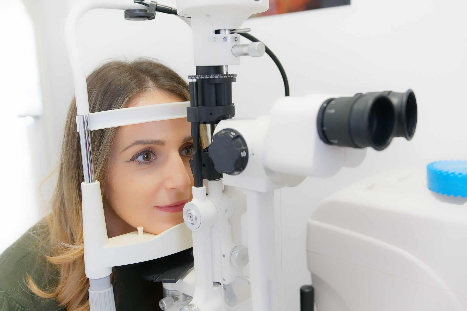
Optometry commonly provides initial vision screening, refraction, and anterior segment evaluation for many blur causes. Ophthalmology involvement is indicated when slit-lamp or fundus examination suggests retinal, optic nerve, or surgical pathology, or when urgent intervention may be needed. Referral may also follow detection of signs such as afferent pupillary defect, significant vision loss, or retinal tears. Coordination between optometry and ophthalmology ensures timely diagnostics and appropriate escalation to subspecialty care, for example retina, neuro-ophthalmology, or glaucoma services.
What are medical and surgical treatments?
Treatment depends on the underlying cause. Medication can address inflammatory or infectious optic neuropathies, intraocular inflammation, and some vascular conditions where systemic management is critical. In cases of retinal vascular occlusion, therapy focuses on managing risk factors and, in selected cases, intravitreal injections for secondary macular edema. Surgery is required for retinal detachment, advanced cataract affecting vision, or urgent decompression in some optic nerve compressive lesions. Therapies should be evidence-based and discussed with the treating clinician, emphasizing risks, expected outcomes, and follow-up requirements.
How do rehabilitation and screening support recovery?
Rehabilitation and ongoing screening aid visual recovery and prevent recurrence. Low-vision rehabilitation provides devices and training for persistent deficits. Visual therapy may help in specific binocular or accommodative disorders. Regular screening for systemic contributors such as diabetes and hypertension reduces risk of recurrent vascular events that affect eyesight. Post-treatment monitoring with scheduled imaging and refraction checks detects complications early and guides rehabilitation intensity. Multidisciplinary care, including occupational therapy when functional vision loss affects activities of daily living, supports patient safety and adaptation.
Sudden visual blur spans a spectrum from benign refractive changes to sight-threatening or systemic emergencies. A structured approach—history, focused examination, targeted refraction, and selective imaging—helps differentiate causes and determine whether urgent intervention or conservative management is appropriate. Clear communication between optometrists, ophthalmologists, and primary care providers improves diagnostic efficiency and aligns treatment with patient-specific needs. Regular screening for systemic risk factors and timely rehabilitation measures support long-term visual function and quality of life.


