Non-surgical strategies for reducing vision cloudiness and glare
Cloudy vision and increased glare can interfere with daily tasks such as driving, reading, or working on screens. Many non-surgical approaches can reduce fogginess and light halos by addressing underlying causes, optimizing optical correction, and improving ocular surface health. This article summarizes common causes, diagnostic steps, and practical non-invasive strategies that clinicians and patients often consider.

What causes vision cloudiness and glare?
Cloudiness and glare are symptoms rather than diagnoses and can arise from multiple sources: tear film instability, corneal irregularities, cataract formation, retinal changes, refractive errors, or neurologic processing differences. Patients often report blur that varies through the day, increased haloes around lights at night, or difficulty with contrast. A thorough symptom history helps prioritize potential causes and guides targeted assessment. Recognizing whether symptoms are intermittent or progressive is important for planning optics, ocular surface care, or further diagnostic testing.
How do refraction and optics affect symptoms?
Uncorrected refractive error, subtle astigmatism, or poor lens centration can create persistent blur and glare. Refraction testing and trial lenses remain first-line, especially when symptoms improve with simple correction changes. Modern optics—such as anti-reflective coatings, blue-light filters, and aspheric lens designs—can reduce perceived glare for some individuals. Proper spectacle or contact lens fitting, including consideration of multifocal designs, often reduces symptom burden without invasive measures. Regular refraction reassessment is advised when symptoms change.
When is ophthalmology evaluation and imaging needed?
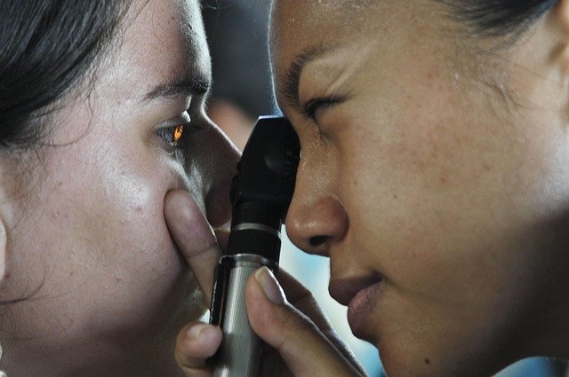
An ophthalmology assessment helps differentiate anterior and posterior causes. Standard evaluations include slit-lamp examination of the cornea and lens, intraocular pressure check, dilated fundus exam, and imaging such as anterior segment photography, optical coherence tomography (OCT), or retinal imaging when macular or retinal disease is suspected. Diagnostic testing clarifies whether corneal disease, early cataract, macular degeneration, or other pathology is present, and it directs non-surgical versus surgical management decisions.
Could neurology or retina issues be responsible?
Neurology and retina conditions may present with clouded vision or photopsias that change with gaze or lighting. Retinal disorders—such as diabetic maculopathy, epiretinal membrane, or early macular degeneration—can reduce contrast and cause distortion where non-surgical rehabilitation or monitoring may be appropriate. Neurologic causes including migraine, optic neuropathies, or cortical visual processing disorders require collaboration with neurology and tailored diagnostic pathways. Symptom pattern and targeted imaging guide these referrals.
What non-surgical cornea and lenses options exist?
Corneal surface problems like dry eye, epithelial irregularities, and mild corneal scarring often respond to topical therapies and optical modifications. Treatments include lubricating drops, anti-inflammatory topical medication for inflammatory surface disease, lid disease management, and therapeutic contact lenses to regularize the ocular surface. Specialty contact lenses—scleral or rigid gas permeable designs—can mask corneal irregularities and reduce glare without surgery. Lens choices and coatings for spectacles also play a role in improving night vision and reducing stray light.
Rehabilitation, medication, and ongoing assessment
Vision rehabilitation strategies complement medical therapy: contrast-enhancing filters, improved lighting, and visual training exercises can help patients adapt. Medications for ocular surface inflammation or tear supplementation can improve optical quality and reduce symptoms. Regular assessment—refraction checks, ocular surface evaluation, and imaging when indicated—ensures timely adjustments. When non-surgical measures provide insufficient relief, findings from these assessments inform whether surgical options may be appropriate in the future.
This article is for informational purposes only and should not be considered medical advice. Please consult a qualified healthcare professional for personalized guidance and treatment.
In summary, reducing vision cloudiness and glare non-surgically involves a stepwise approach: identify likely causes through symptom assessment and targeted diagnostics, optimize optics with appropriate refraction and lens choices, treat ocular surface and retinal contributors when present, and employ rehabilitation techniques to improve functional vision. Coordination between primary eye care providers, ophthalmology, and other specialists helps ensure that strategies are tailored to each patient’s clinical picture.


