Reducing biofilm recolonization after subgingival cleaning
Effective management of biofilm recolonization after subgingival cleaning helps support gingival health and reduce inflammation. This article outlines practical clinical and home strategies to limit plaque return, promote healing, and support the oral microbiome.
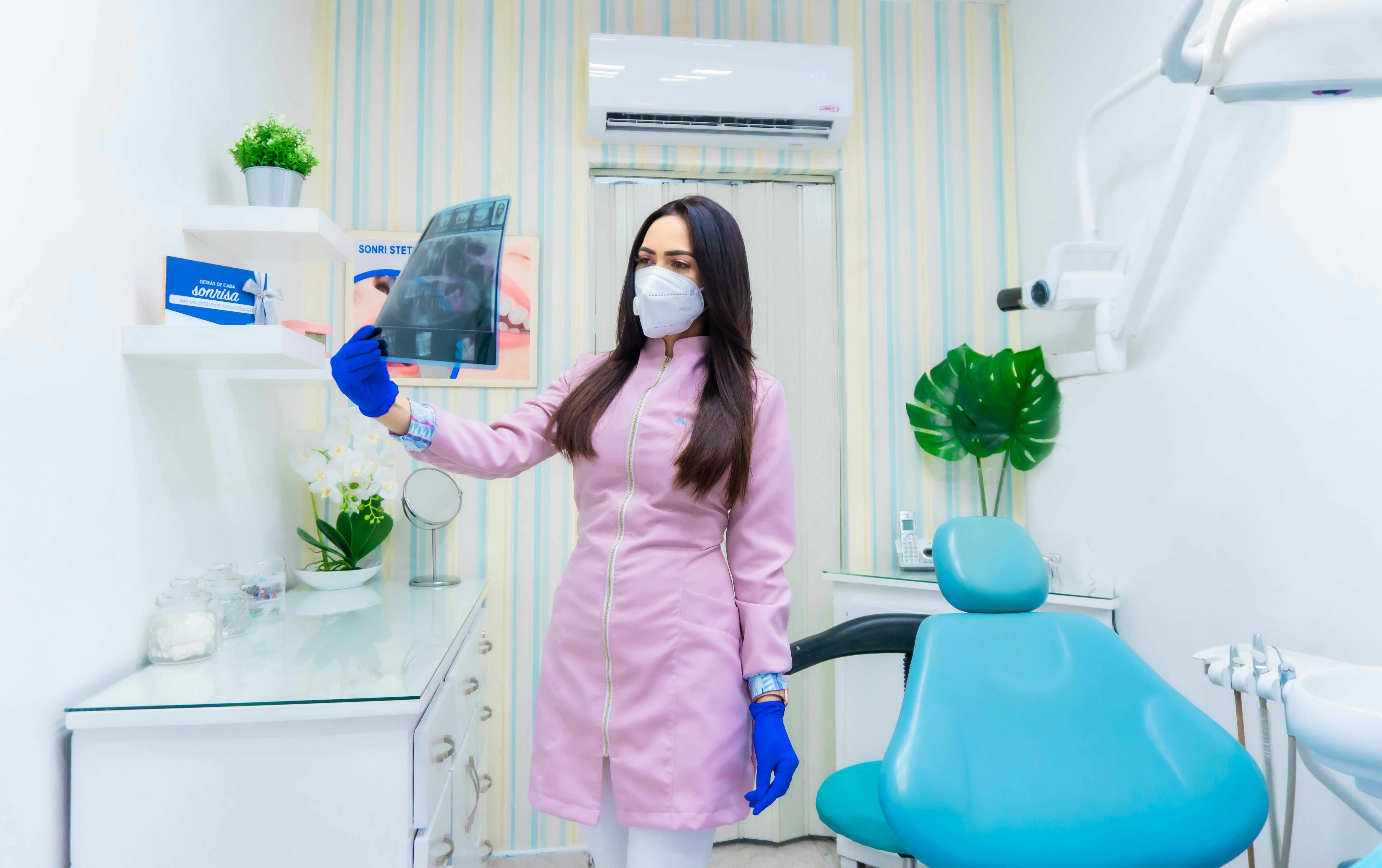
Successful subgingival cleaning – whether by scaling and root planing or professional debridement with adjunctive tools – removes established plaque and calculus from periodontal pockets but does not permanently prevent recolonization. Biofilm communities are resilient: bacteria left on root surfaces, in deep pockets, on the tongue, and in interdental spaces can repopulate cleaned sites within days to weeks, reestablishing a microbial challenge for the gingiva. Effective post‑treatment management therefore combines well‑executed mechanical therapy, targeted adjuncts, tailored maintenance schedules, and patient behavior changes to reduce the pace and impact of recolonization while supporting tissue healing and minimizing sensitivity. Understanding how biofilm forms, how the oral microbiome responds to disruption, and which monitoring and prevention measures are practical in clinical practice and at home helps clinicians and patients set realistic expectations and sustain periodontal stability.
This article is for informational purposes only and should not be considered medical advice. Please consult a qualified healthcare professional for personalized guidance and treatment.
Gingiva and biofilm: how recolonization occurs
The gingiva is the primary tissue affected when biofilm re‑establishes after subgingival therapy. Remaining plaque and microbial reservoirs in pocket niches allow pathogenic species to regain foothold, prolonging local inflammation and slowing healing. Mechanical removal reduces the bulk of biofilm but the root surface and pocket topography can retain microcolonies. The host response of the gingiva—ranging from mild hyperemia to persistent inflammation—depends on the balance of plaque load, tissue susceptibility, and systemic factors. Regular assessment of gingival status following treatment helps identify early recolonization before more extensive tissue changes occur.
Role of the oral microbiome in recolonization
The oral microbiome comprises diverse bacteria that respond dynamically to disruption. After debridement, rapid shifts occur: early colonizers repopulate the space first, forming a biofilm scaffold that later permits more complex species to join. Maintaining a balanced microbial community that favors health‑associated species over pathogenic shifts is an objective of many maintenance strategies. Measures that reduce overall plaque and disrupt biofilm architecture can tilt recolonization toward less inflammatory microbial profiles while supporting ecological healing in the treated pocket.
Impact of scaling, debridement, and healing
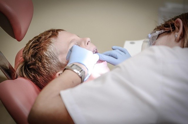
Scaling and subgingival debridement reduce pathogenic load and facilitate clinical attachment reformation and pocket depth reduction where possible. Healing after these interventions includes re‑epithelialization and connective tissue reattachment, processes that are sensitive to ongoing microbial challenge. Controlled debridement that minimizes unnecessary trauma supports faster repair and reduces post‑procedure sensitivity. Proper technique, adequate time for healing, and scheduled reassessment are important to detect and manage early recolonization before irreversible changes such as recession occur.
Managing interdental plaque and sensitivity
Interdental areas are common reservoirs for plaque and can act as continuous sources of recolonization. Interdental cleaning with floss, interdental brushes sized to the patient’s embrasures, or other aids reduces microbial load between teeth and limits migration into pockets. Patients may experience transient sensitivity after deep periodontal procedures; desensitizing agents, gentle brushing techniques, and avoiding extremely hot or cold foods help during the healing window. Clinicians should instruct patients on interdental technique and select tools that balance efficacy with comfort to encourage adherence.
Prevention, maintenance, and monitoring strategies
A structured maintenance plan is central to preventing rapid recolonization. Short‑interval monitoring visits in the early post‑treatment months allow plaque control reinforcement, selected professional maintenance, and timely interventions if recolonization is detected. Adjunctive measures—such as targeted antiseptics, localized antimicrobial delivery when appropriate, and optimization of oral hygiene devices—can be integrated based on individual risk. Maintenance frequency should be individualized by disease severity, response to therapy, and patient behavior to sustain pocket health and minimize recurrence.
Behavior, inflammation, recession, and long-term outcomes
Patient behavior strongly influences long‑term results. Smoking, poor oral hygiene, and inconsistent attendance for maintenance accelerate recolonization and perpetuate inflammation, increasing the risk of recession and attachment loss. Controlling systemic contributors, encouraging consistent hygiene routines, and educating patients about plaque‑driven disease mechanisms help reduce inflammatory burden. Over time, stable management of biofilm and inflammation supports favorable periodontal outcomes, while unmanaged recolonization can lead to progressive tissue breakdown.
A coordinated approach that integrates meticulous initial debridement, protection of healing tissues, interdental plaque control, individualized maintenance intervals, and patient‑centered behavior change reduces the likelihood and clinical impact of biofilm recolonization. Monitoring and adapting strategies according to the patient’s response preserves tissue health and supports long‑term stability without relying on any single intervention.

