Referral Pathways: When Primary Care Sends Patients to Specialists
Primary care clinicians often identify skin or subcutaneous bumps, nodules or masses during routine visits. This article outlines typical triggers for referral, how initial evaluation proceeds, and what patients can expect when their doctor sends them to a specialist for further diagnosis and care.

Referral Pathways: When Primary Care Sends Patients to Specialists
Primary care clinicians commonly encounter bumps, nodules, masses or other growths on or under the skin during routine exams or when patients report symptoms. When a lump raises concern—because of pain, rapid growth, changes in skin appearance, or worrisome features on an exam—the clinician will weigh monitoring against further testing and possible referral. This first step balances the likelihood that a finding is benign with the need to exclude malignant or serious causes, using history, focused examination, and initial diagnostic choices to guide the pathway.
This article is for informational purposes only and should not be considered medical advice. Please consult a qualified healthcare professional for personalized guidance and treatment.
What symptoms prompt a referral?
Primary care physicians consider several symptoms when deciding whether to refer a patient for specialist assessment. Red flags include lumps that are growing quickly, painful, fixed to underlying tissue, associated with skin ulceration, unexplained weight loss, or systemic symptoms such as fever or night sweats. Location and context matter: masses deep in muscle or subcutaneous tissue, rapidly enlarging nodules on the torso, or lumps near lymph node basins often prompt earlier referral. Persistent or recurrent lesions despite initial treatment also merit specialist input to clarify diagnosis and next steps.
How do primary care doctors begin diagnosis?
Initial diagnosis focuses on history and targeted physical exam: onset, progression, associated symptoms, prior trauma, infection history, and family or personal cancer history. Clinicians may perform basic office investigations such as measuring the size, assessing mobility, and palpating for tenderness. Laboratory tests or empiric short-term treatment can be used to rule out inflammatory or infectious causes. If the clinical picture is unclear, or if a mass has concerning features, primary care providers often order imaging or arrange referral rather than delay definitive evaluation.
When is biopsy recommended?
Biopsy is recommended when tissue diagnosis is necessary to distinguish benign from malignant processes or to guide specific treatment. Indications include masses with suspicious imaging features, unexplained subcutaneous nodules that persist or grow, and lesions where infection or inflammatory causes have been excluded. A specialist—dermatologist, general surgeon, or oncologic surgeon—will select the biopsy type (excisional, incisional, core needle, or punch) based on size, location, and likelihood of malignancy. Biopsy provides histopathology that informs staging, prognosis, and treatment choices.
How are imaging tests used?
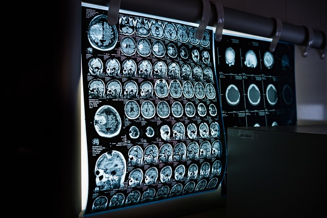
Imaging helps define the size, depth, and internal character of bumps, nodules, or masses. Ultrasound is a common first-line modality for superficial masses, offering information about cystic versus solid nature and vascularity. MRI is used when deeper tissue definition is needed or when soft-tissue tumor characterization and surgical planning are required. Plain X-rays can identify calcifications or bone involvement. Imaging often guides whether a lesion can be monitored, needs biopsy, or requires surgical referral for removal.
How can patients selfcheck and monitor lumps?
Patients are encouraged to perform regular, gentle selfchecks and to note changes in size, shape, mobility, color, or associated symptoms like pain or discharge. Monitoring includes photographing a lesion over time and recording dates of any changes. Primary care clinicians may recommend a defined observation period with scheduled followup if a lesion appears benign. Clear instructions on red-flag changes that warrant urgent reassessment—rapid growth, new pain, skin breakdown, or systemic symptoms—help ensure timely referral when needed.
What happens after referral: treatment and followup?
Referral routes depend on the suspected diagnosis: dermatologists often evaluate superficial skin and subcutaneous lesions, general surgeons handle larger soft-tissue masses or excisions, and oncologists manage confirmed malignancies. Treatment options range from continued monitoring to surgical excision, targeted therapies, or systemic treatment for malignant disease. Followup plans are tailored to pathology results and risk: benign excised lesions may need minimal monitoring; atypical or malignant findings require structured surveillance with periodic imaging, clinical review, and coordination across specialties to manage recurrence risk and long-term outcomes.
Patients benefit from clear communication between primary care and specialists, with shared records of imaging, biopsy results, and treatment plans. This collaborative approach helps ensure that bumps, nodules, masses or growths are accurately classified and managed according to clinical risk.
In summary, referrals from primary care to specialists are guided by symptom patterns, clinical suspicion from examination, and results from initial diagnostic steps such as ultrasound or biopsy. Understanding these referral pathways helps patients anticipate the evaluation process and supports timely, appropriate care for both benign and potentially malignant conditions.


