Rehabilitation approaches after transient loss of sight detail
Transient reductions in visual detail can be alarming. This article outlines practical rehabilitation approaches for people who experience temporary blurred or altered vision, focusing on diagnostic steps, rehabilitation strategies, workplace adjustments, and follow-up care to support recovery and functional clarity.

Transient reductions in visual detail—episodes of blurred vision, increased haze, or intermittent glare—often prompt immediate concern. Understanding the typical course and rehabilitation options helps patients and clinicians prioritize diagnostics, safety, and recovery goals. Rehabilitation after a transient loss of sight detail combines targeted assessments, optical adjustments, workplace ergonomics, medication review, and coordinated follow-up to restore functional clarity and reduce recurrence.
Symptoms: clarity, haze and glare
Many people report a sudden drop in clarity, perceived haze across the visual field, or episodes of glare that make contrast and fine detail difficult to see. Symptoms may be monocular or binocular and can fluctuate with lighting, fatigue, or head position. Noting whether the change affects central detail, peripheral awareness, color perception, or reading ability helps guide assessments. Keep a simple symptom log noting duration, triggers, and recovery time—this information improves diagnostics and subsequent rehabilitation planning.
Diagnostics: imaging, refraction and screening
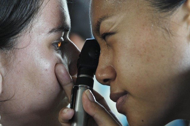
Initial diagnostics typically include visual acuity testing, objective refraction, intraocular pressure checks, and a slit-lamp examination of the cornea and anterior segment. Imaging such as optical coherence tomography (OCT) can assess retina and optic nerve health, while fluorescein angiography or fundus photography may be used when vascular or retinal pathology is suspected. Neurological screening, including pupil reflex testing and visual field checks, identifies patterns consistent with neurological or vascular causes. A coordinated screening pathway reduces missed diagnoses and informs tailored rehabilitation.
Retina and cornea: structural considerations
Structural problems in the retina or cornea are common contributors to temporary visual detail loss. Corneal irregularities, dry eye, or epithelial disturbances produce haze and reduce clarity at the front of the eye, while macular ischemia, edema, or detachment affect fine central detail at the retina. Rehabilitation often begins with resolving treatable anterior segment issues (lubrication, surface therapy) and managing retinal conditions through specialist referral when imaging suggests structural involvement. Clear differentiation between corneal and retinal origins is essential for effective management.
Neurology and vascular causes to consider
Transient loss of detail can reflect transient ischemic events, migraine-related visual disturbance, or cranial nerve dysfunction. Neurology input is important when symptoms are sudden, recurrent, or accompanied by other neurological signs such as weakness, speech changes, or severe headache. Vascular risk factors—hypertension, diabetes, hyperlipidemia—should be optimized as part of a prevention-focused rehabilitation plan. Collaboration between ophthalmology, neurology, and primary care supports comprehensive evaluation and reduces the risk of recurrence.
Medication, ergonomics and workplace adjustments
Medication side effects and systemic drugs can contribute to blurred vision or glare; review of current medication is a routine step. Ergonomics and lighting in the workplace influence visibility—improved task lighting, antiglare screens, and correct monitor height reduce strain and enhance perceived clarity. Environmental adjustments such as contrast-enhancing materials, reducing reflective surfaces, and scheduled visual breaks support recovery. Occupational therapists or workplace health services can recommend individualized adjustments for those returning to visually demanding tasks.
Rehabilitation strategies and followup plans
Rehabilitation integrates optics (updated refraction, filters, or low-vision aids), surface and ocular health treatments, and graded return-to-activity plans. When optical correction is needed, precise refraction and trial of tints or polarizing lenses can lessen glare and improve detail. Vision rehabilitation specialists can introduce visual training exercises focused on saccadic accuracy and contrast sensitivity where appropriate. Followup schedules vary by cause but often include short-term re-evaluation within days to weeks and longer-term imaging or neurologic review if symptoms persist or recur. Clear documentation and patient education on warning signs ensure timely re-presentation if new symptoms arise.
This article is for informational purposes only and should not be considered medical advice. Please consult a qualified healthcare professional for personalized guidance and treatment.
Conclusion A measured approach to rehabilitation after transient loss of sight detail emphasizes accurate diagnostics, correction of reversible optical or surface problems, interdisciplinary assessment for neurological or vascular contributors, and practical workplace and lifestyle adjustments. Structured followup and individualized rehabilitation plans improve functional outcomes and help people regain visual clarity while reducing the risk of future episodes.


