Stepwise clinical assessment for acute visual blurring
Acute visual blurring requires a structured clinical approach to identify reversible causes and urgent threats to sight or life. This brief overview outlines key steps clinicians use to evaluate sudden blur, including history, basic examination, targeted investigations, and early management considerations.

Acute visual blurring can arise from many causes, ranging from refractive changes to retinal or neurological emergencies. A stepwise clinical assessment helps determine whether immediate intervention is needed and guides appropriate referral. The initial evaluation prioritizes patient safety, distinguishes monocular from binocular blur, and identifies symptoms that suggest urgent imaging or specialist input. Timely, systematic assessment reduces diagnostic delay and supports targeted treatment, rehabilitation, or monitoring options.
How is visual acuity assessed?
Visual acuity is the foundational test in any assessment of vision and blur. Measure distance and near acuity using standardized charts or portable equivalents. Record results with habitual correction, then test using pinhole to screen for significant refractive error versus media opacity. Document whether blur is constant or intermittent and whether acuity improves with pinhole or corrected lenses. Accurate acuity measurement guides decisions about refraction, optical correction, and urgency of further evaluation.
When is refraction checked?
Refraction is essential when blurred vision could be optical rather than pathological. Perform objective (autorefractor or retinoscopy) and subjective refraction if pinhole or history suggests refractive error. Sudden onset blur in younger patients may follow contact lens issues or abrupt accommodation changes; in older adults, uncorrected hyperopia or cataract progression can present as subacute blur. Correcting refractive error with glasses or contacts can often restore vision, while lack of improvement directs clinicians to inspect the cornea, lens, and retina.
How are cornea, lens, and retina examined?
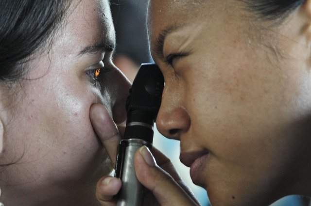
A focused anterior and posterior segment exam evaluates the cornea, lens, and retina for causes of blur. Slit-lamp or torch examination can reveal corneal abrasions, edema, or infections; lens opacity suggests cataract; fundus exam assesses macular pathology, retinal hemorrhages, detachment, or vascular occlusions. Portable ophthalmoscopy and dilated fundoscopy improve visualization. Findings such as a relative afferent pupillary defect, macular changes, or peripheral retinal tears inform urgency and specialty referral.
What imaging and diagnosis steps are used?
When clinical exam is limited or neurological signs exist, imaging becomes important. Optical coherence tomography (OCT) provides detailed retinal and macular assessment. Fundus photography and fluorescein angiography help evaluate retinal circulation and lesions. Neuroimaging (MRI or CT) is indicated when visual blur is accompanied by headache, diplopia, field defects, altered mental status, or signs suggesting optic nerve or intracranial pathology. Proper selection of imaging modalities supports accurate diagnosis and management planning.
When is neurology and medication considered?
Neurological causes of acute blur—optic neuritis, ischemic optic neuropathy, stroke affecting visual pathways—require timely neurology involvement. Medication history is critical: some drugs can induce visual disturbance or affect accommodation. Systemic conditions such as giant cell arteritis, autoimmune disease, or diabetes may present with vision changes and need coordinated medical therapy. Early identification enables prompt administration of steroids, anticoagulation, or other targeted medication when indicated by diagnosis.
What rehabilitation, contacts, and telemedicine options exist?
After acute management, rehabilitation and low-vision support assist patients with persistent deficits. Contact lens issues are managed with hygiene education, lens replacement, or alternative corrections. Telemedicine can be useful for triage, follow-up, or remote monitoring of visual acuity and symptoms, especially for local services or patients with access barriers. Referral for vision rehabilitation, occupational therapy, or community services helps restore function and quality of life when permanent changes occur.
This article is for informational purposes only and should not be considered medical advice. Please consult a qualified healthcare professional for personalized guidance and treatment.
Clinical assessment of acute visual blurring follows a logical sequence: measure acuity, assess refraction and anterior segment, perform fundus evaluation, and select imaging or specialist referral as indicated. Clear documentation of symptom onset, associated signs, and response to simple interventions (pinhole, refractive correction) directs appropriate next steps. Integrating ophthalmic and medical perspectives—alongside rehabilitation and telemedicine options when appropriate—supports comprehensive care and more timely diagnosis.


