Understanding Macular Degeneration: Causes, Signs, Care
Macular degeneration is a top cause of central vision loss in adults over 50. Learn how the macula is affected, what early signs to watch for, how clinicians diagnose it, and the latest treatments and lifestyle steps that can slow progression and preserve sight. Discover practical prevention tips and support options.

Macular degeneration damages the macula, the small central area of the retina responsible for sharp, detailed vision. While peripheral sight usually remains intact, damage to this tiny but vital region makes reading, recognizing faces, driving, and other near tasks increasingly difficult. Detecting changes early and working with eye care professionals can help preserve usable vision and quality of life.
How macular degeneration alters vision
The condition primarily compromises central vision. In its initial stages, people often notice mild blurring or subtle distortions when focusing straight ahead. As the disease progresses, central details grow less distinct and a gray or dark patch may develop in the center of the visual field. There are two principal forms:
- Dry (atrophic) macular degeneration: This more common type advances gradually, driven by thinning of retinal tissue and accumulation of drusen—tiny yellow deposits beneath the retina.
- Wet (neovascular) macular degeneration: Less common but potentially severe, this form involves abnormal blood vessel growth beneath the retina that can leak fluid or blood and cause rapid vision loss.
Understanding which form is present guides monitoring and treatment choices.
Early signs to monitor
Catching symptoms early allows timely intervention. Key warning signs include:
- Mild central blurriness, especially when reading
- Difficulty seeing small print or fine detail
- Colors that look duller or less vivid than before
- Straight lines appearing wavy or bent
- A small blurry or dark spot in the middle of your vision
If these changes appear suddenly or worsen quickly, contact an eye care professional without delay.
Risk factors and likely contributors
Macular degeneration arises from a mix of genetic, environmental, and health-related factors. Known risk factors include:
- Age: Risk increases substantially after age 50.
- Family history and genetic variants: Certain genes raise vulnerability.
- Smoking: A major modifiable risk that accelerates progression.
- Ethnicity: Higher prevalence among people of Caucasian descent.
- Obesity and poor cardiovascular health: These can hasten disease.
- High blood pressure and cholesterol: Associated with greater risk.
- Diet low in antioxidants or high in saturated fat.
- Long-term ultraviolet light exposure.
While age and genetics can’t be changed, addressing modifiable risks can slow decline.
How clinicians diagnose macular degeneration
Routine eye exams are crucial, particularly for adults over 50 or anyone with risk factors. Common diagnostic steps include:
- Visual acuity testing to check clarity of vision
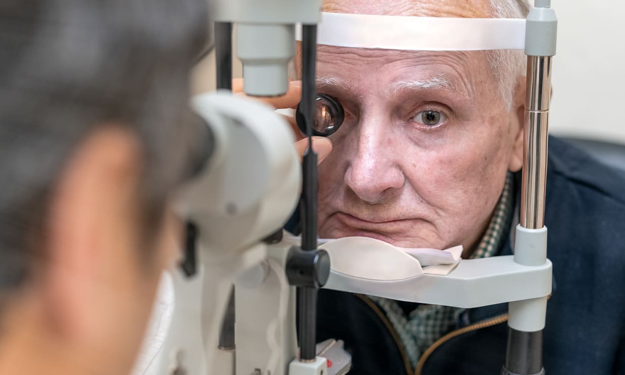
- Dilated eye exams to directly view the retina and macula
- Amsler grid testing at home to detect distortion or central blind spots
- Optical coherence tomography (OCT) to create cross-sectional images of retinal layers and identify fluid accumulation
- Fluorescein angiography to map leaking or abnormal blood vessels in suspected wet AMD
These evaluations help classify the stage and type of macular degeneration and inform treatment planning.
Treatment approaches
Although there is no cure, several therapies can slow progression or manage symptoms:
- Anti-VEGF injections: For wet AMD, medications injected into the eye block vascular endothelial growth factor, reducing abnormal vessel growth and fluid leakage.
- Photodynamic therapy: A light-activated drug combined with laser treatment can target leaking vessels in select cases of wet AMD.
- Laser photocoagulation: In limited scenarios, laser can seal problematic blood vessels.
- Nutritional supplements: AREDS and AREDS2 formulations (specific combinations of vitamins and minerals) have been shown to lower the risk of progression in certain intermediate or late-stage dry AMD cases.
- Lifestyle measures: Quitting smoking, eating leafy greens and omega-3-rich fish, exercising, and controlling blood pressure and cholesterol support eye health.
- Low vision aids: Magnifiers, specialized lighting, screen readers, and adaptive technologies help people maintain independence despite central vision loss.
| Treatment | Purpose |
|---|---|
| Anti-VEGF injections | Reduce abnormal blood vessel growth and leakage in wet AMD |
| Photodynamic therapy | Seal leaking vessels using a light-activated drug and laser |
| Laser photocoagulation | Target and seal specific leaking vessels in some cases |
| AREDS / AREDS2 supplements | Nutritional support to slow progression in certain dry AMD stages |
| Low vision aids | Improve daily function through magnification and adaptive tech |
Cost disclaimer: Treatment costs vary by provider, location, and insurance; consult your healthcare provider or insurer for specific pricing information.
Slowing progression and prevention
While you can’t modify age or genetics, several actions can reduce risk or slow disease progression:
- Stop smoking and avoid secondhand smoke.
- Eat a nutrient-rich diet with leafy greens, colorful vegetables, and oily fish.
- Maintain a healthy weight and stay physically active.
- Manage blood pressure and cholesterol through lifestyle and medications as needed.
- Wear sunglasses that block UV rays and use wide-brimmed hats outdoors.
- Keep up with regular eye exams and use an Amsler grid at home to spot new changes promptly.
- Talk with your eye doctor about whether AREDS or AREDS2 supplements are appropriate for your stage of disease.
Living with macular degeneration
Adjusting to central vision loss can be both practical and emotional. Vision rehabilitation specialists and occupational therapists can teach strategies and recommend devices that maximize remaining vision. Home modifications, adaptive technology, smartphone apps, and magnification tools can preserve independence. Support groups, counseling, and family communication also help with the emotional impact.
Ongoing contact with your eye care team is essential: quick evaluation of sudden changes, adherence to treatment schedules (for example, timely anti-VEGF injections), and proactive lifestyle measures all contribute to better long-term outcomes.
This article is for informational purposes only and should not be considered medical advice. Please consult a qualified healthcare professional for personalized guidance and treatment.


